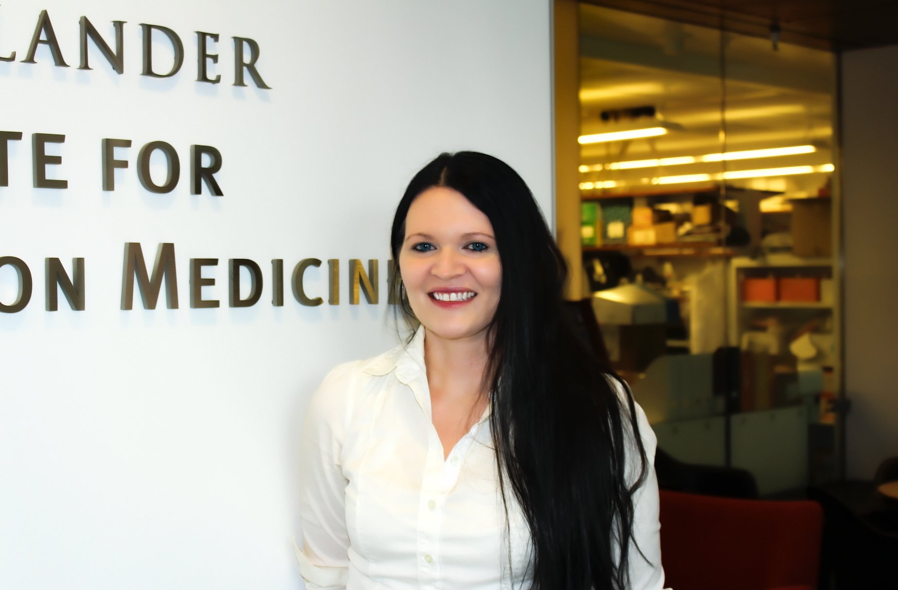
We are pleased to introduce our colleague Jenna Moyer, a Research Specialist on the EIPM organoid team.
We hope you enjoy learning more about her research interests and background!
Question: Can you please tell me about your work with organoids and how you first learned about this area of research?
A: I have been involved with organoid development and research applications for almost six years. I first began optimizing protocols to derive organoid cancer models under a National Cancer Institute initiative to create a repository of cell line models to be accessible to researchers. Upon moving to NYC, I joined the EIPM organoid platform. At EIPM, we generate organoids from patient tumors which we then characterize molecularly, screen with our high throughput drug screening robot, and use in research applications focused on understanding cancer and finding potential therapeutics.
Q: What are some of the challenges you regularly face and overcome in this capacity?
A: Cancer is a very complex disease and, even within one tissue type, such as endometrial cancer, each case is different because each person is unique. Therefore, each organoid model is different and must be developed and maintained in the lab while keeping those nuances in mind. Just as there is no “one-size fits all” treatment for cancer, there is not one broadly applicable protocol for easily establishing organoids from every tissue we receive. We continue to optimize the individually required factors and conditions needed for long-term growth of each organoid culture so that it can be expanded for multiple screens and assays.
Q: How important are organoids to the field of precision medicine and patient care?
A: Organoids are 3-dimensional cell line models that can form tissue-like structures which mimic the actual tissue from where they are derived. This makes them ideal for laboratory driven research investigating patient-specific tumor development and therapeutics. Organoids can be analyzed genetically for cancer driving mutations and screened against hundreds of therapeutics. This can provide clinicians and researchers with personalized treatment insights for patient care.
Q: Where do you see the next technological advances coming for your work?
A: Along with increasingly efficient drug-screening robotics, high content imaging has great potential for evaluating therapeutics. Currently, EIPM is optimizing our drug screening protocols to integrate data from a 3D imager which can evaluate more than 50 parameters, such as cellular morphology and texture. Organoids can be imaged and evaluated throughout the entire course of drug treatment. Because this imaging is non-invasive and does not utilize any endpoint chemical assays, the cells can be kept in culture for subsequent second line treatment screens and so on.
Another advancing aspect of organoid modelling is the bioengineering of tissue specific scaffolding to support the 3-dimensional structures. Organoids are 3D because they are grown in a gel-like substance that keeps them suspended. This allows the cells to self-organize and develop into specialized structures without the interference of laboratory 2D surfaces. These gels are often made from structural proteins that support cells and tissues within the body. By engineering more accurate scaffolds, the organoids can grow in environments much closer to their origin location, resulting in more accurate research applications.
Q: What is the most satisfying part of your work?
A: It is satisfying to be part of a team with such driven ideals and ambitious ideas. Everyone, from clinicians to researchers to collaborators to donors, all have a common goal: to further cancer research in order to save lives. Being part of such a collaborative environment is what moves science forward.
Q: How has the Covid-19 pandemic affected your work?
A: I joined EIPM during the beginning of the pandemic. We did not want to lose 3D models under development, so I quickly joined the other staff on a rotation schedule to maintain laboratory operations. Although research applications had to slow for a time, we continued to research, organize, and learn. Using video calls to communicate with clinicians and other scientists, we continued to work on essential projects. Video meetings really enable us to meet other researchers virtually to share ideas, plans, and discussion. Although the laboratory is now fully staffed, we continue to meet virtually with collaborators across the institution and around the world.
Q: What keeps you motivated?
A: Each model we develop and characterize adds to our knowledge about cancer. Everything we learn puts us one step closer to finding the next successful treatments for cancer patients. Working so closely with clinicians who provide a bridge between patient and research reminds me every day of why I am a scientist and who I am researching for. There is purpose in knowing that each sample received and each model developed will have lasting impacts on the field of cancer research.
Q: What do you like to do outside of work?
A: Out of the lab, I do a lot of reading and writing, mostly science fiction. I also like hiking (or city-walking) and rowing, which allow me to push myself physically while enjoying a bit of peaceful meditation.
# # #
