The Englander Institute for Precision Medicine strives to be at the forefront of translational research. We’ve recently paired the Helios™, mass cytometry by time-of-flight (CyTOF) instrument with the Hyperion imaging module. The system is one of 30 of its kind in use, and the only one in the NYC area.
With this advanced technology for single-cell analysis, we are providing investigators with a powerful, affordable and actionable tool for exploring complex biological processes.
Imaging Mass Cytometry (Fluidigm Hyperion)
Imaging Mass Cytometry (Fluidigm Hyperion)
The Fluidigm Hyperion imaging mass cytometry (IMC) system allows highly multiplexed tissue imaging, currently at up to ~40 markers per panel using metal-tagged antibodies and at 1 µm/pixel resolution for images comparable to light microscopy with minimal spillover between channels. This technology enables high-content, deep profiling of complex functional and signaling states of various cells in the tissue immune microenvironment (TIME) with spatial and structural context, unveiling the important interactions between these cells and their tissue and disease context.
Helios (Single Cell Suspension)
CyTOF Mass Cytometry (Fluidigm Helios)
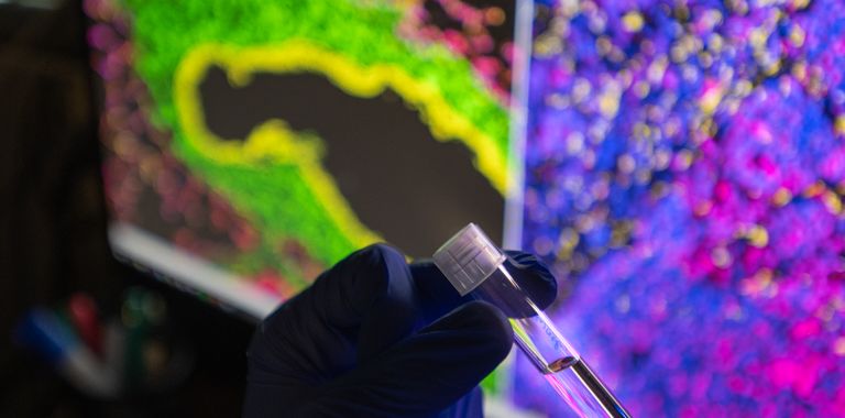
High parameter spatially resolved protein detection at sub-cellular resolution using mass cytometry time of flight in situ (CyTOF Hyperion).

Resources and Publications
- The underlying principles and emerging applications explained
- Mass Cytometry Publications
- CyTOF and Helios Publications